A New Direction in Restoring Interproximal Caries in Posterior Teeth: The Bioclear Method
Since then, considerable innovative ideas have been implemented into merging the old with the new. During this process, we have learned that composite restorations are susceptible to fracture, staining, sensitivity, poor interproximal contacts, recurrent decay, overhanging margins, voids and thus shorter longevity than their amalgam counterparts¹. Many gadgets and techniques have developed over the years to help handle composite resins predictably using traditional metal matrix bands. In an era when people are more aware of esthetics and decry the use of amalgam², manufacturers have been obsessed with improving the chemistry and placement technique of composite resins to overcome their shortcomings.
There is another variable to this equation. What if we, similarly, modernized cavity preparations to shapes that would take advantage of the mechanical properties of this material to make stronger, predictable, longer-lasting class 2 restorations?
Invented by Dr. David Clark, Bioclear matrices and the Bioclear Method are replacing traditional G.V. Black Class 2 restorations with a modern, science-proven procedure that aims to strengthen and prolong the service life of posterior composites. The perfect marriage between material and technique could potentially improve the longevity of our composites, especially for endodontically-treated teeth that would be weakened by a heavy cervical chamfer preparation to support a crown. There are five pillars to the Bioclear Method that need to be followed systematically to engineer strength and durability into interproximal posterior composite restorations.
There are five pillars to the Bioclear Method that need to be followed:
1. Biofilm Removal
Isolation with a rubber dam is facilitated by light interproximal sanding with a fine IPR strip (Bioclear TruContact) and gently opening up any occlusal embrasures with a coarse disc in order to avoid accidental tearing of the rubber dam on sharp enamel or flat marginal ridges upon placement. Teeth are covered in plaque. Always. And some of the microorganisms that live in it contribute to the degradation of resin restorations³. Removal of biofilm provides a clean surface for the adhesion of composite resins, which in turn reduces staining and recurrent caries at the tooth-restoration interface⁴. Liberally applying a disclosing solution, Bioclear Dual-Colour Disclosing Solution, followed by removal with Bioclear Blasting Powder (Al₂OH₃ powder in a high-pressure Blaster (Bioclear), provides a clean surface for bonding (Fig. 1, 2 and 3). The aluminum tri-hydroxide powder removes biofilm safely without damaging tooth structure.
2. Preparation Design
The Clark Class 2 preparation is designed to minimize stress concentration, to facilitate the flow of warm composite resin and to increase the surface area of enamel bonding. Boxes and bevels are no longer needed, and it is no longer necessary to cut dentin to create mechanical retention. The square corners and sharp lines of G.V. Black preparations are unfavourable for composite bonding. Polymerization contraction forces (C-factor) increase with the number of bonded surfaces of a cavity preparation⁵. A ratio greater than 3:1 of bonded to non-bonded surfaces is considered unfavourable, leading to the formation of gaps at the interface between tooth and resin, and new gaps will continue to form over time ⁶,⁷,⁸,⁹.
A Bioclear Diamond Wedge is ideal for pre-wedging due to its 2 lbs of separation force, its expanding leading point that locks it into place and its robust spine that will endure bur contact. Once the pre-wedge is in place, initial access to caries is made with a tapered diamond bur leaving the contact intact. The contact is now broken toward the buccal and lingual using a blunt needle diamond bur, and the preparation is extended into an infinity margin several millimeters onto the buccal and lingual surfaces. After removing the pre-wedge, a safe-sided finishing strip further blends the margin and softens any edges. The occlusal cavo-surface margin is similarly flared. Designing broad, smooth radius walls on enamel increase the surface area for bonding and minimize shrinkage stress. Dentin is instrumented only to remove decay (Fig. 4 and 5). For endodontically-treated teeth, the preparation is engineered so that there is cuspal coverage, a long-standing principle that significantly prolongs the service life of the tooth¹⁰.
3. Anatomic Tooth Forms (Bioclear Matrices)
Bioclear matrices mimic the natural anatomy of anterior and posterior teeth. They come in different shapes and heights to accommodate different clinical scenarios. Selection is streamlined by measuring the height of the proximal box and comparing this value against the BioFit Matrix Selection Guide.
These matrices can be customized by the clinician to match the restorative needs of the tooth. They are transparent to facilitate curing of the resin and are stiffer than metal matrices. The pre-curved natural interproximal contacts and embrasures eliminate the need for burnishing (Fig. 6), however inserting the placement plier and expanding the tips bucco- and lingually will stretch the matrix, ensuring a broad contact.
By using the radiograph as a guide to inter-root space and relative crestal bone height to select the appropriate size of wedge, a second and larger Bioclear Diamond Wedge is placed while firmly holding down the folded occlusal tab to stabilize the matrix. The available Deep Caries wedge is ideal for the deep subgingival margin placed in the furca. Placement of the separating ring completes the Bioclear Twin Ring Sectional Matrix System.
4. Injection-molded Heated Composite
Heating paste and flowable composites lowers their viscosity and enables better adaptation to the cavity preparation and reduces the incidence of voids¹¹,¹². Heating composite has no detrimental effect on the pulp or the mechanical, esthetic and light-curing properties of the material and final restoration¹³. These claims are made specifically for 3M composites as this company has rigorously tested their materials for compatibility with heat.
Once the preparation has been etched, rinsed and its adhesive light-cured, a second application of adhesive is applied, air-thinned but not polymerized. Flowable composite, heated in a HeatSync (Bioclear) along with two loaded composite guns to 155°F (68°C) is injected in between the matrix and the tooth, following the U-shape of the preparation. Immediately the heated paste composite is injected into the uncured flowable and adhesive allowing the backpressure to gently lift the tip out of the tooth until the entire preparation is filled beyond the occlusal cavo-surface margin. If necessary, the other heated syringe is used to complete this injection molding technique. At no time does the tip of the compule leave the material during injection. Excess composite is removed with a Curved Sculpting Paddle (Bioclear) or a Sculpting Point (Bioclear) and gentle primary anatomy is established. Three-point light-curing (occlusal, buccal and lingual) ensures complete polymerization due to the transparency of the matrix.
The objective of injecting composite is to bulk-fill the tooth to create a monobloc that gets sculpted back to a final shape (Fig. 7); a technique that simplifies the filling procedure and provides strength to the restoration¹⁴,¹⁵.
5. Rockstar Polish
A high-gloss finish reduces the formation of bacterial biofilm on the surfaces of composite restorations¹⁶. Using coarse and/or medium grit Sof-Lex™ (3M) discs and the proprietary MagicMix (Bioclear) followed by RS Diamond Polishers (Bioclear), one can achieve a mylar-like finish to direct composites (Fig. 8 and 9). This mylar-like finish reduces the formation of bacterial biofilm on the surface of the new restoration, helping prolong its longevity¹⁷.
Summary
Dentistry will continue to restore interproximal caries for the foreseeable future. Manufacturer innovation will continue to improve the magic that drips out of dental adhesive bottles and extrudes out of composite compules. Following systematically the aforementioned protocol will help prolong the service life of teeth that would have been extracted otherwise. The Bioclear Method, when properly applied, can make a difference in your clinical practice and the health of your patients.
Interested in learning more about the Bioclear Method?
Clinical Technique

FIG. 1
Bioclear Dual-Colour Disclosing Solution is applied to the tooth to be restored as well as the adjacent teeth.
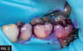
FIG. 2
After 5 – 10 seconds, the disclosing solution is rinsed off, revealing the location of the biofilm.
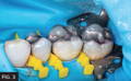
FIG. 3
Using a Bioclear Blaster with aluminum tri-hydroxide, the biofilm is removed. Pre-wedging with Bioclear Diamond Wedges is established to protect the rubber dam and gingival papilla from accidental bur damage.
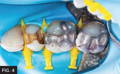
FIG. 4
The old amalgam restoration is removed revealing sharp edges from the G.V. Black preparation.
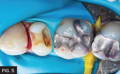
FIG. 5
The Bioclear non-retentive preparation is complete. Note that all sharp edges are removed and the dentin has been “blasted” with aluminum tri-hydroxide.
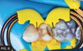
FIG. 6
BioFit matrices, Diamond wedges and TwinRing separating rings are in place.
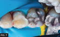
FIG. 7
Preparation has been over-filled with 3M Filtek Supreme Ultra with the injection molding technique.
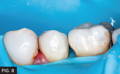
FIG. 8
Preparation has been over-filled with 3M Filtek Supreme Ultra with the injection molding technique.
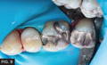
FIG. 9
Final restoration with Rock Star polish.
REFERENCES
- Bernardo M, Luis H, Martin MD, Leroux BG, Rue T, Leitão J, DeRouen TA. Survival and reasons for failure of amalgam versus composite posterior restorations placed in a randomized clinical trial. Am. Dent Assoc. 2007 Jun;138(6):775-83. doi: 10.14219/jada.archive.2007.0265. PMID: 17545266.
- https://www.mercuryconvention.org/Portals/11/documents/Booklets/COP3-version/Minamata-Convention-booklet-Sep2019-EN.pdf
- Pinna R, Usai P, Filigheddu E, Garcia-Godoy F, Milia E. The role of adhesive materials and oral biofilm in the failure of adhesive resin restorations. Am Dent. 2017 Oct;30(5):285-292. PMID: 29178733.
- Jacker-Guhr S, Sander J, Luehrs AK. How "Universal" is Adhesion? Shear Bond Strength of Multi-mode Adhesives to Enamel and Dentin. Adhes Dent. 2019;21(1):87-95. doi: 10.3290/j.jad.a41974. PMID: 30799475.
- Carvalho RM, Pereira JC, Yoshiyama M, Pashley DH. A review of polymerization contraction: the influence of stress development versus stress relief. Oper Dent. 1996 Jan-Feb;21(1):17-24. PMID: 8957911.
- Okuda M, Pereira PN, Nakajima M, Tagami J, Pashley DH. Long-term durability of resin dentin interface: nanoleakage vs. microtensile bond strength. Oper Dent. 2002 May-Jun;27(3):289-96. PMID: 12022462.
- Braga RR, Ferracane JL, Condon JR. Polymerization contraction stress in dual-cure cements and its effect on interfacial integrity of bonded inlays. Dent. 2002 Sep-Nov;30(7-8):333-40. doi: 10.1016/s0300-5712(02)00047-7. PMID: 12554115.
- Yoshikawa T, Sano H, Burrow MF, Tagami J, Pashley DH. Effects of dentin depth and cavity configuration on bond strength. Dent Res. 1999 Apr;78(4):898-905. doi: 10.1177/00220345990780041001. PMID: 10326734.
- Roulet JF. Marginal integrity: clinical significance. J Dent. 1994;22 Suppl 1:S9-12. doi: 10.1016/0300-5712(94)90164-3. PMID: 8201087.
- Aquilino SA, Caplan DJ. Relationship between crown placement and the survival of endodontically treated teeth. JProsthet Dent. 2002 Mar;87(3):256-63. doi: 10.1067/mpr.2002.122014. PMID: 11941351.
- Fróes-Salgado NR, Silva LM, Kawano Y, Francci C, Reis A, Loguercio AD. Composite pre-heating: effects on marginal adaptation, degree of conversion and mechanical properties. Dent Mater. 2010 Sep;26(9):908-14. doi: 10.1016/j.dental.2010.03.023. Epub 2010 Jun 16. PMID: 20557926.
- Lucey S, Lynch CD, Ray NJ, Burke FM, Hannigan A. Effect of pre-heating on the viscosity and microhardness of a resin composite. Oral Rehabil. 2010 Apr;37(4):278-82. doi: 10.1111/j.1365-2842.2009.02045.x. Epub 2009 Dec 29. PMID: 20050987.
- Lopes LCP, Terada RSS, Tsuzuki FM, Giannini M, Hirata R. Heating and preheating of dental restorative materials-a systematic review. Clin Oral Investig. 2020 Dec;24(12):4225-4235. doi: 10.1007/s00784-020-03637-2. Epub 2020 Oct 20. PMID: 33083851.
- Rosatto CM, Bicalho AA, Veríssimo C, Bragança GF, Rodrigues MP, Tantbirojn D, Versluis A, Soares CJ. Mechanical properties, shrinkage stress, cuspal strain and fracture resistance of molars restored with bulk-fill composites and incremental filling technique. Dent. 2015 Dec;43(12):1519-28. doi: 10.1016/j.jdent.2015.09.007. Epub 2015 Oct 9. PMID: 26449641.
- Versluis A, Douglas WH, Cross M, Sakaguchi RL. Does an incremental filling technique reduce polymerization shrinkage stresses? Dent Res. 1996 Mar;75(3):871-8. doi: 10.1177/00220345960750030301. PMID: 8675797.
- Versluis A, Douglas WH, Cross M, Sakaguchi RL. Does an incremental filling technique reduce polymerization shrinkage stresses? Dent Res. 1996 Mar;75(3):871-8. doi: 10.1177/00220345960750030301. PMID: 8675797.
- Kurt A, Cilingir A, Bilmenoglu C, Topcuoglu N, Kulekci G. Effect of different polishing techniques for composite resin materials on surface properties and bacterial biofilm formation. J Dent. 2019 Nov;90:103199. doi: 10.1016/j.jdent.2019.103199. Epub 2019 Sep 23. PMID: 31557551.
About the Author

Rafael A. Bustamante, BSC, DDS
Rafael Bustamante graduated from the University of Lethbridge in 2000 with a degree in Biochemistry and received his dental degree from the University of Alberta in 2004. His interests revolve around the application of the dental operating microscope in his daily procedures; a technology he adopted upon graduating from dental school. Doing restorative dentistry under high magnification has taken him on a journey to strive daily for perfection and exactness.
Discover More
This article was originally published in the Clinical Life™ magazine: Spring 2021 edition
Clinical Life™ magazine is a premier periodical publication by Clinical Research Dental Supplies & Services Inc. Discover compelling clinical cases from Canadian and US dental professionals, cutting-edge techniques, product insights, and continuing education events.
Subscribe to our emails to receive articles like this and be notified about our exclusive promotions.