Case Study: Clinical Management of Deep Subgingival Margin Elevation in Posterior Teeth with Structural Compromise
The understanding of restorative dentistry today demands not only considering the preservation of remaining tissues but also respecting the biomechanics governing the dental structures, having as main objectives the preservation of pulpal health and strengthening fragile and more affected teeth, providing a clinical treatment with the longest possible lifespan.
Within routine clinical practice, restorative concepts and techniques aim to bioemulate lost tissues in terms of structure and function. Precisely one of the most
demanded techniques today for our indirect restorative procedures is the relocation of cervical margins or deep margin elevation. This clinical approach was first described in 1998 by Dietschi and Spreafico, who defined it as “a technique indicated in cases with slightly subgingival margins, where it is possible to relocate
the cervical area of a preparation more coronally by applying appropriate increments of composite resin on the existing margin. This procedure should be developed under absolute isolation with a rubber dam, followed by the placement of a metal matrix band” (1). Since the introduction of this new concept, other authors and publications have emerged, emulating the technique, and applying it to various scenarios in restorative dentistry. In terms of posterior indirect partial restorations, elevating the cervical margin of a preparation above the gingival margin provides advantages for impression taking and subsequent isolation during the cementation process. However, fundamental requirements must be taken into consideration in order to ensure a correct adhesive procedure: complete isolation of the cervical margin of the preparation and placement of a matrix band that achieves perfect sealing of the cervical area. Otherwise, the technique is contraindicated.
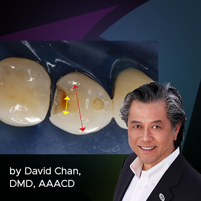
Once the technique is performed, a bitewing radiograph should be taken to evaluate the adaptation of the composite resin in the gingival area, checking for gaps
or over-contours before proceeding with the final impression (2). In terms of clinical elements to consider for correct clinical performance, a curved matrix is recommended to ensure not only perfect sealing in the gingival area but also the formation of a correct emergence profile. In this situation, the use of ReelMatrix™ Deep Margin Elevation Bands from Garrison is recommended. (Fig. 1)
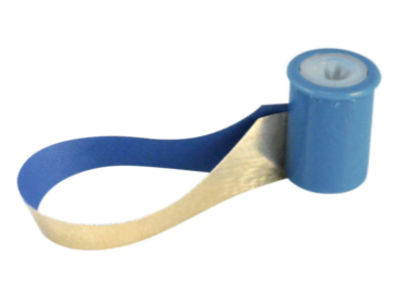
FIG. 1
Due to their thickness, contour, and height, these bands achieve anatomical margin elevation in most cases. Like other bands designed by the same company, they
have two different surfaces: one coated with Teflon to improve handling of the restorative material and another with a rigid metallic surface.
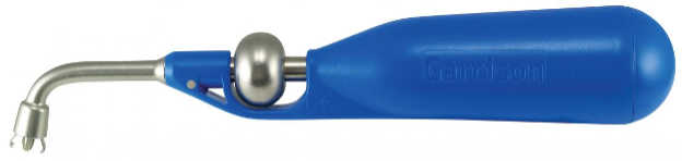
FIG. 2
This band, which was specially designed for this technique, should be attached using a tensioning instrument (Fig. 2) which places this band on the cervical margin of the preparation with efficiency and precision.
Clinical Case
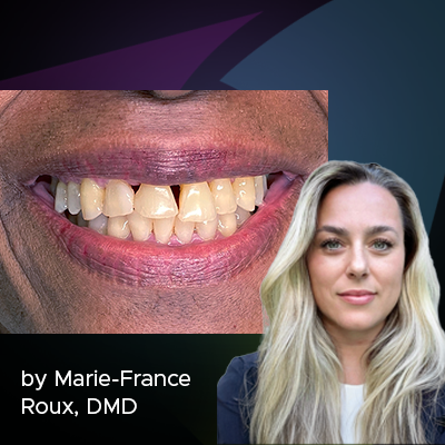
A 20-year-old female patient visits the dental clinic due to moderate pain and expresses a desire to replace an old restoration. Upon clinical examination, a composite
resin restoration partially covering the occlusal surface of tooth 2.4 is observed, showing marginal gaps, secondary caries, and deficient anatomy (Fig. 3).
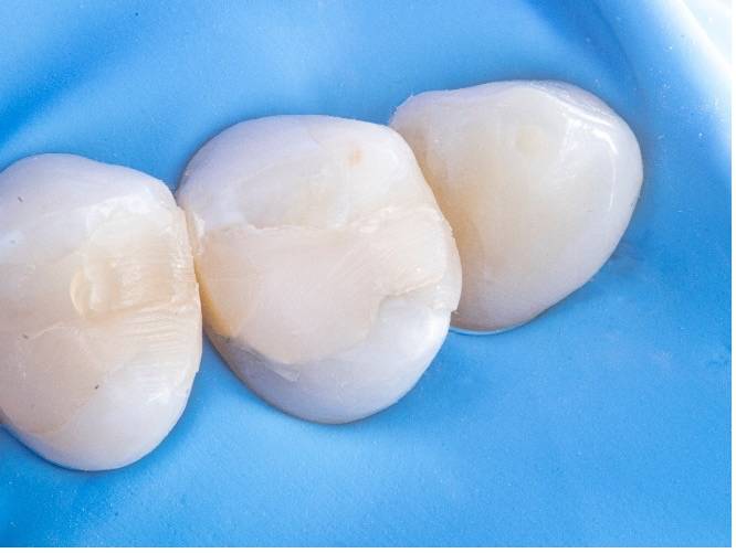
FIG. 3
Based on the specific structural diagnosis, an indirect composite resin restoration on a stress-controlled biobase was indicated. This will maximize adhesive strength through a rigorous biomimetic restorative protocol associated with deep margin elevation in the mesial proximal box without the need for prior surgical intervention.
The procedure described above was performed in two phases. The first phase focused on primarily replacing the dentin, for which the old restoration was completely
removed (Fig. 4).
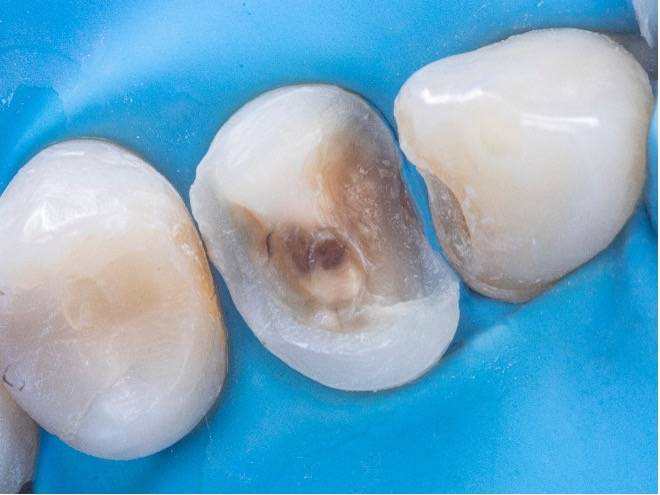
FIG. 4
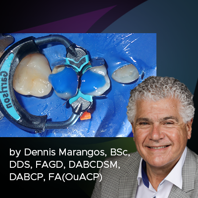
Subsequently, the tensor-band complex was placed to elevate the cervical margin using the ReelMatrix™ Deep Margin Elevation system (Fig. 5).
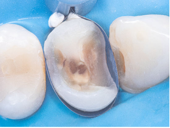
FIG. 5
The image shows the band’s adaptation to the tooth, achieving a perfect marginal seal (Fig. 6).
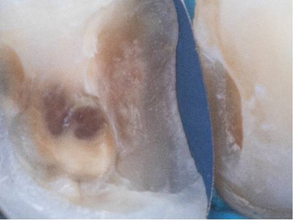
FIG. 6
Immediately after, the adhesive phase was completed using Clearfil SE Bond 2 by Kuraray, followed by the application of a hydrophobic layer of Grandioso Heavy
Flow by Voco and finally a 3 mm strip of Ribbond polyethylene fiber to ensure adhesion, along with a thin layer of high-load microhybrid resin, in this case, Clearfil APX
by Kuraray (Fig. 7).
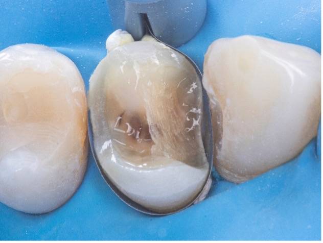
FIG. 7
Subsequently, layers of composite resin, not exceeding 1mm in thickness, were applied to achieve progressive buildup. Once the biobase was established, a minimal
biological preparation was performed using diamond burs, and a definitive impression was taken for the fabrication of the indirect restoration (Fig. 8).
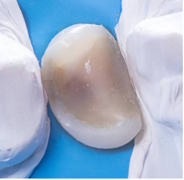
FIG. 8
In the second phase, under absolute isolation, the indirect restoration was connected to the previously constructed biobase using an adhesive bonding agent, in this case, a heated composite resin, to ensure an extended working time, high mechanical properties, and favorable aesthetics. Excess material was carefully removed, mainly in proximal areas, and after curing, the rubber dam was removed for the final view of the restoration, evaluating harmony with the periodontal tissue and occlusion (Fig. 9).
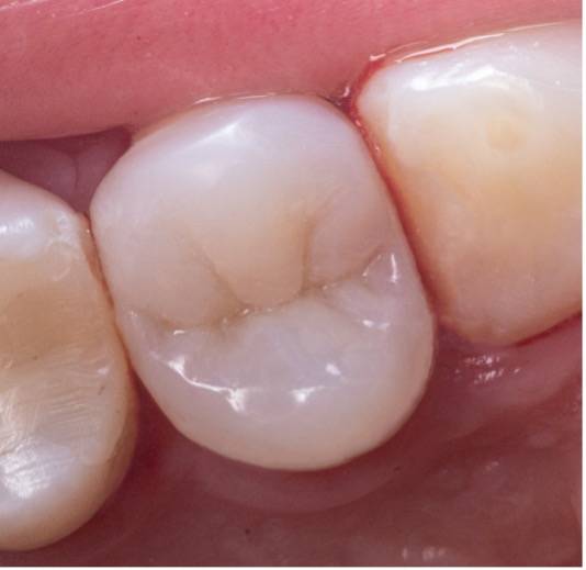
FIG. 9
REFERENCES
1) D. Dietschi et al. Current clinical concepts for adhesive cementation of tooth - colored posterior restoration. Pract Periodontal Aesthete Dent (1998) 47–54.
2) P. Magne, R. Spreafico. Deep Margin Elevation: A paradigm Shift Am J Esthet Dent 2012;2:86–96.
©2023 Garrison, Kuraray, Voco and Ribbond. All rights reserved.
All other trade names referenced are the service marks, trademarks or registered trademarks of their respective companies.
About the Author
Patricio Gutiérrez Pino, DDS
Dr. Patricio Gutiérrez Pino is a graduate of the dental school of Pedro de Valdivia University, Santiago, Chile. He has carried out his entire career in the private sector practicing in various clinics located in Santiago. Since receiving his degree, Dr. Gutiérrez Pino has trained and practiced restorative and aesthetic dentistry as this is his area of interest. In 2016, he received the aesthetics diploma in adhesive oral rehabilitation at the dental school of the University of Los Andes, Santiago. After that, he held teaching positions at universities and institutes contributing numerous publications and clinical cases. This vocation led him to become a Magister in Higher Education at Andres Bello University. Currently, Dr. Gutiérrez Pino is studying to obtain the Diploma in Restorative Dentistry Biomimetics at Diego Portales University in Santiago. In recent years, Dr Gutiérrez Pino has dedicated himself exclusively to this area and to becoming a Key Opinion Leader of different commercial dental brands that are established in Chile.
Discover More
This article was originally published in the Clinical Life™ magazine: Fall 2023 edition
Clinical Life™ magazine is a premier periodical publication by Clinical Research Dental Supplies & Services Inc. Discover compelling clinical cases from Canadian and US dental professionals, cutting-edge techniques, product insights, and continuing education events.
Subscribe to our emails to receive articles like this and be notified about our exclusive promotions.