Gingival Sculpting With a Soft Tissue Diode Laser
With today’s selfie culture, more and more patients are requesting improved or idealized cosmetics. Often times what appears to be small changes can give big results and happy patients.
Evaluating the gingival architecture is an important component of every smile design. When the gingival zeniths are asymmetric across the midline, the smile can appear unpleasing to the eye. Performing gingival sculpting will enhance the smile. Gingivectomy is a surgical procedure of excising some of the free gingival tissue to create a new, more apical gingival margin. It is imperative to be mindful of the biological width when considering gingival sculpting. The biologic width should not be violated. Biologic width is about 2mm which consists of about 1mm of epithelial tissue and 1mm connective tissue attachment. Encroaching on the biologic width may cause gingival recession or gingival rebound and compromise the health of the tissue. Maintaining an adequate zone of keratinized tissue must also be considered.
This case illustrates a common cosmetic concern I see in my practice. “I want my smile to look better. I’ve whitened my teeth and I still don’t love the way my teeth look.” This patient has very healthy and nicely shaped teeth but is dissatisfied with their appearance. Smile evaluation showed she had a black triangle between her central incisors. She also asymmetry of her gingival with her left central and lateral incisors having lower gingival margins and zeniths than her right central and laterals. (FIG. 1)
Simple and conservative procedures can get her the results she desires without the need for removing tooth structure for more extreme measures like veneers. The patient had more tooth whitening in-office. The black triangle was closed with direct composite bonding. Additionally, gingival sculpting was performed with the Bluewave diode laser (Clinician’s Choice). (FIG. 2)
Gingivectomy can be performed with several different methods. In the past, I have used a scalpel and also electrosurge. Both have disadvantages including shrinkage of tissue during healing. The diode laser is a good choice. Diode laser is highly absorbable by hemoglobin and melanin that allows easy manipulation of soft tissue during gingival recontouring, and improved epithelization and healing of the wound. There is no bleeding and minimal pain during the procedure. The tissue healing is very predictable with no shrinkage. These, along with precise control, are all benefits of using a diode laser for soft tissue surgery. Precise placement of the margin can be accomplished. (FIG. 3)
The Bluewave diode laser is a great choice. It is powerful and simple to use. It is easily portable from operatory to operatory. I like the magnetic handpiece rest.
Soft tissue laser surgery has some disadvantages that include the cost, buying a laser device is expensive comparing it to scalpel. The Bluewave diode is at an affordable price point and can be used for many soft tissue laser procedures.
The one week post-op photo shows the improved smile note the healing of the gingiva at the precise position of the laser surgery. (FIG. 4) These conservative procedures achieved the esthetic results the patient desired while being mindful of her long-term dental health.
Clinical Case
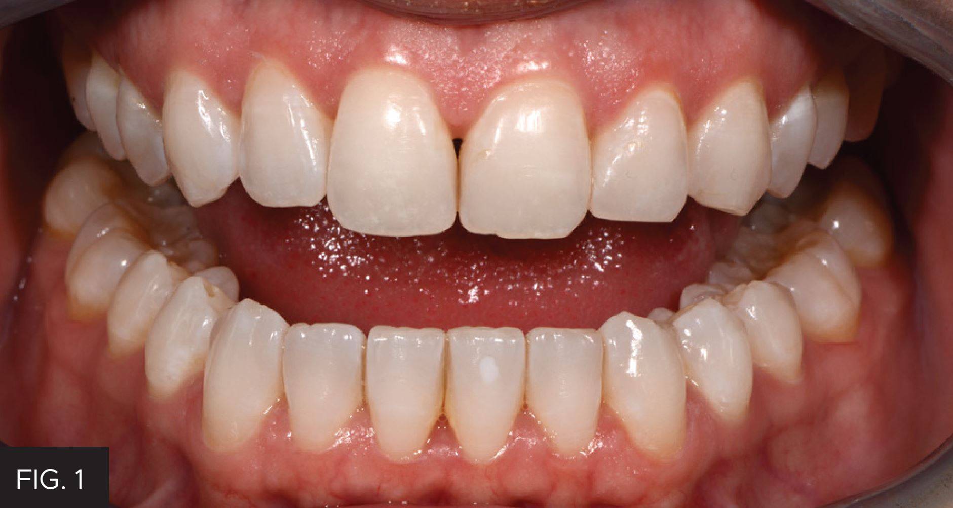
FIG. 1
Pre-op. Note black triangle between the central incisors and the asymmetry of the gingiva with lower gingival margins on the left central and lateral incisor.
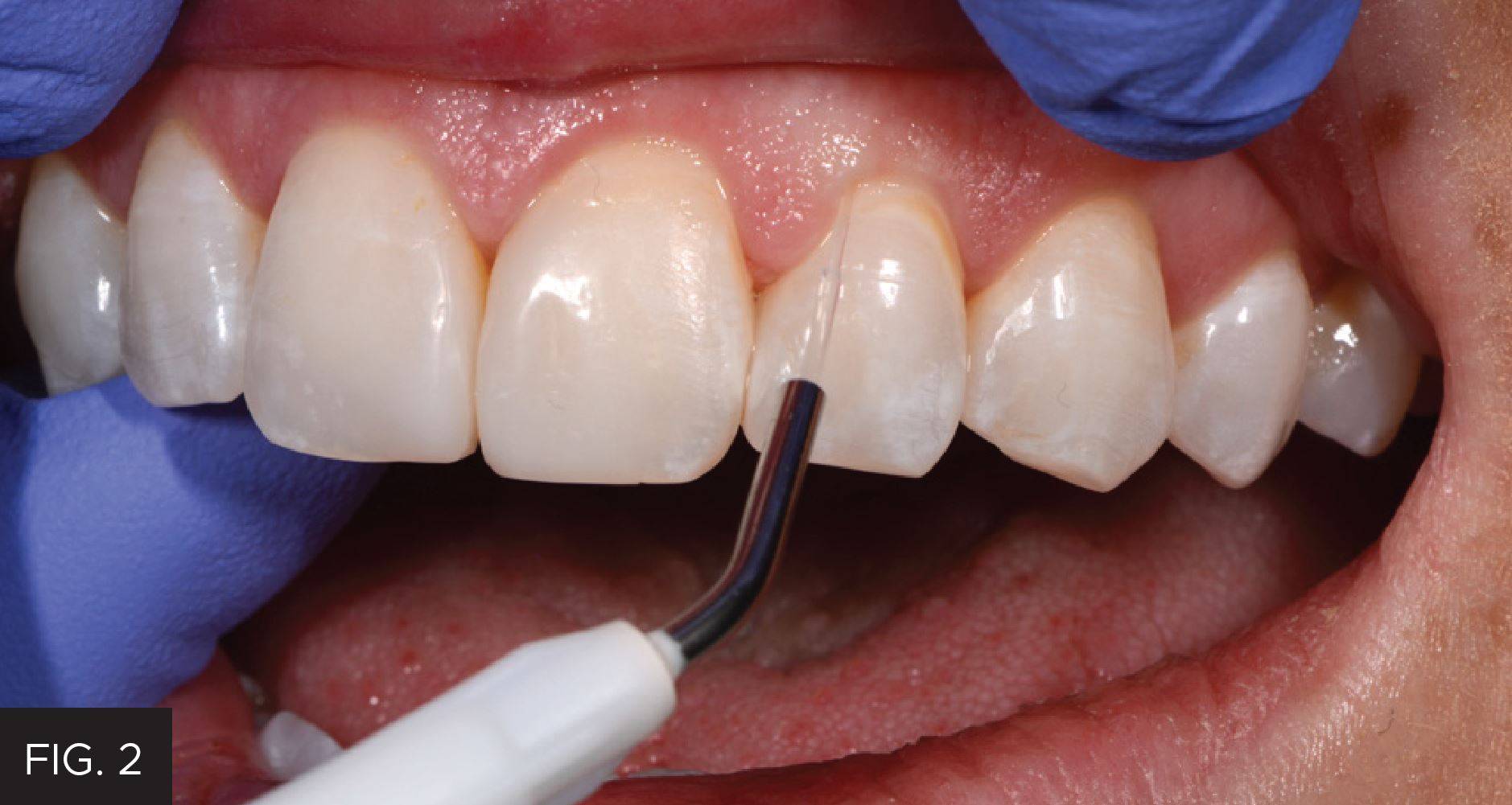
FIG. 2
After whitening and black triangle closure, gingival sculpting is performed with the Bluewave diode laser.
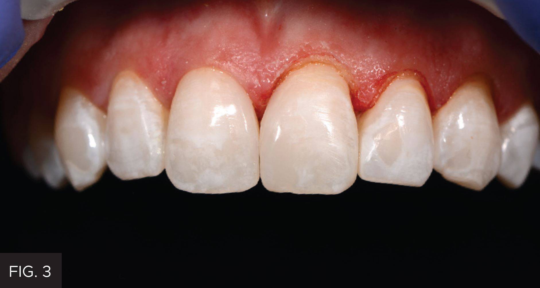
FIG. 3
After following my bonding protocol, we were able to place our final restoration in a stress-free environment due to the Bluewave controlling a very difficult situation. (FIG. 4)
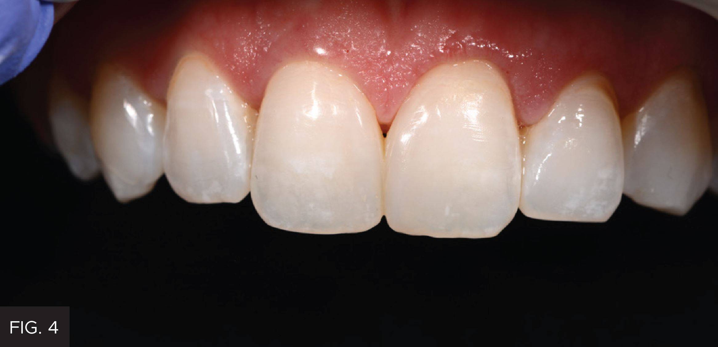
FIG. 4
One week post-op. Symmetric gingival with margins at exact position of laser surgery.
Images courtesy of Susan McMahon DMD, AAACD, AGD
About the Author

Susan McMahon, DMD, AAACD, AGD
Dr. McMahon enjoys one of the largest cosmetic dental practices in Western Pennsylvania. She is accredited by the American Academy of Cosmetic Dentistry, and a Fellow of the prestigious American Society for Dental Aesthetics and a Fellow in the Academy of General Dentistry. An author and lecturer, Dr. McMahon has devoted her professional career to the pursuit of advanced technologies in cosmetic and minimally invasive dentistry. She is the Director of New Product Evaluation for Catapult Education. She frequently lectures across the United States on minimally invasive dentistry, technology and conservative cosmetics. She has been voted by her peers as a Top Pittsburgh Dentist every year for over 20 years.
Discover More
This article was originally published in the Clinical Life™ magazine: Spring 2022 edition
Clinical Life™ magazine is a premier periodical publication by Clinical Research Dental Supplies & Services Inc. Discover compelling clinical cases from Canadian and US dental professionals, cutting-edge techniques, product insights, and continuing education events.
Subscribe to our emails to receive articles like this and be notified about our exclusive promotions.